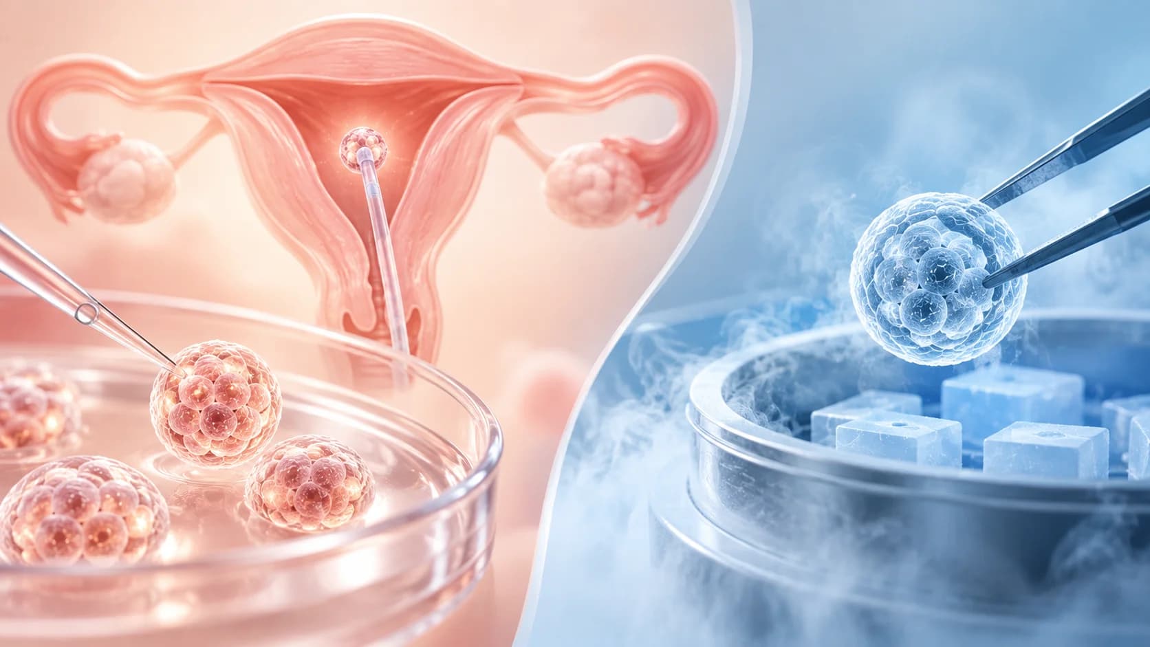
New-Generation Technologies That Improve IVF Success

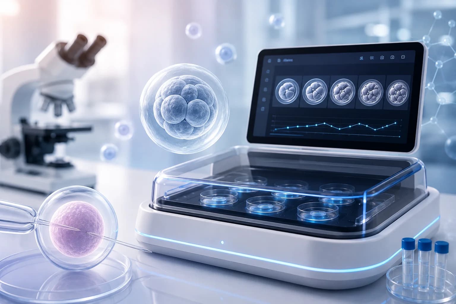
1. EMBRYOSCOPE (Continuous Monitoring System):
Success in IVF treatment is not the result of science alone, but of the right use of advanced technologies that track even the smallest detail. To offer our patients world-class care and sustain our success rates, which keep rising year after year, we closely follow every innovation with proven reliability. In line with this vision, we are proud to be the first center in Bursa to successfully use the EmbryoScope technology, which has revolutionized how embryo development is followed. With the EmbryoScope, a new era begins in IVF treatment: we no longer focus only on snapshots, but on the embryo's entire life story.
The EmbryoScope is essentially a highly advanced "smart incubator". It allows embryos to grow in conditions that closely mimic the mother's womb, while a special built-in camera records them around the clock.
Here is how it differs from the conventional method: Normally, checking on embryos means taking them out of their warm, safe home, even if only briefly. With the EmbryoScope, the lid is never opened, so the balance of temperature, light and humidity is never disturbed. While our future babies rest peacefully, we watch over them from the outside.
EmbryoScope (Time-Lapse) Technology in IVF:
The most critical step that determines success in IVF (in vitro fertilization) treatment is choosing the right embryo. One of the most important innovations to transform this process in recent years, the EmbryoScope (Time-Lapse Imaging - TLI), makes it possible to monitor embryo development "without interruption". But does this technology really improve the chance of success? Let's look at the advantages and the clinical realities of the EmbryoScope in light of the latest scientific evidence.
What Is the EmbryoScope (Continuous Monitoring)? How Does It Work?
It is an advanced incubator (smart incubator) system that allows embryos developing in the embryology laboratory to be watched moment by moment while being kept in conditions as close as possible to the mother's womb. Thanks to its built-in camera system, a photo is taken every 10-20 minutes without the embryo ever being moved, and the whole development process is recorded like a film strip.
In the conventional method, embryos are taken out of the incubator (an artificial womb environment) once a day and examined under a microscope to check their development. The EmbryoScope, however, thanks to its built-in camera, offers:
- Uninterrupted Monitoring: Photos of the embryos are taken automatically every 10-20 minutes and followed through a computer system.
- A Stable Environment: Because the embryos are not exposed to the outside environment, temperature, pH and gas balance remain undisturbed.
- Morphokinetic Data: Not only the embryo's appearance at the moment of checking, but also dynamic data such as the speed and timing of cell divisions (morphokinetic parameters) are analyzed.
Why Should Embryos Not Be Disturbed?
In the laboratory, an embryo is extremely sensitive to the outside world. The "uninterrupted culture" advantage highlighted in the literature matters for three critical reasons:
- Temperature and Gas Balance: The environment the embryos live in (pH and temperature) must mimic the mother's womb. Every time the incubator lid is opened, this delicate balance is disturbed. With the EmbryoScope, the lid is never opened and the environment always stays stable.
- Reducing Stress Factors: Exposure to light and room air can cause oxidative stress in developing cells. Leaving embryos undisturbed allows them to divide "in peace", without exposure to these external factors.
- Not Missing Developmental "Moments": Some developmental errors (for example, a cell dividing incorrectly and then correcting itself) can be missed during one-off checks. Continuous monitoring without disturbing the embryo allows us to capture these critical moments.
- In short: In conventional methods, embryo development is checked by taking the embryos out once a day and looking at them under a microscope. Yet the embryo is extremely sensitive to the outside world. The greatest advantage the EmbryoScope offers is an uninterrupted culture environment. This stable environment lets embryos grow in safer conditions and increases live birth rates.
Advantages of the EmbryoScope: Why Choose It?
Recent studies in the literature show that continuous monitoring technology makes a difference particularly in the following areas:
- Less Subjectivity: It minimizes differences in observation between embryologists and, thanks to algorithms, provides a more objective assessment.
- Detecting Anomalies: Abnormal divisions that could be missed with a conventional microscope (dynamic anomalies such as dividing directly into three cells) are easily detected with this system.
- Compatibility with Genetic Testing (PGT-A): The EmbryoScope can optimize the process by predicting in advance which embryos are suitable for biopsy. It helps determine the right timing for the biopsy.
- Workflow Efficiency: It increases the safety of embryos in the laboratory while allowing their development to be recorded digitally.
Choosing the Highest-Quality Embryo: Morphokinetic Assessment
The EmbryoScope is not just a monitoring device; it is also an analysis tool that helps us choose the healthiest embryo. While classical methods look only at the embryo's shape at a given moment, the morphokinetic assessment used at our center also analyzes the timing and speed of the embryo's cell divisions.
It should be remembered, however, that using the EmbryoScope requires considerable expertise and experience. Our team, highly experienced in this advanced method that is very different from classical assessments, analyzes EmbryoScope data with great care and selects the embryo with the highest chance of achieving a pregnancy.
The EmbryoScope has opened a new era in IVF treatment. At the first center in Bursa to use this technology, guided by scientific evidence and in expert hands, you too can move one step closer to your dreams.
Scientific Facts: Does It Improve Success for Everyone?
Although the EmbryoScope is a revolutionary device, clinical research emphasizes that it does not miraculously increase live birth rates in every patient.
- A Personalized Approach: It can help by providing more data, especially in specific groups such as patients with recurrent IVF failure or advanced maternal age.
- Live Birth Rates: Meta-analyses have not always found a dramatic difference in live birth rates between standard methods and the EmbryoScope. This shows that the technology is meaningful not on its own, but when combined with laboratory quality and expertise.
What Does the Future Hold?
The EmbryoScope has become the "eye" of IVF laboratories. In the future, when combined with artificial intelligence (AI) integration and non-invasive biomarkers (which do not harm the embryo), its potential to identify the healthiest embryo will grow even further.
In short, the EmbryoScope is not just a monitoring device, but a technology that records the embryo's life story. It is important to discuss in detail with your doctor whether this technology is right for you during your treatment.
The EmbryoScope gives embryologists a kind of "time machine". It lets us look not only at the embryo's current shape, but at its entire developmental story. This improves our chance of choosing the right embryo and brings you one step closer to your dreams.

Monitoring with an EmbryoScope device in the IVF laboratory
2. PGT-A and NGS: Reducing Miscarriage Risk by Selecting a Healthy Embryo
For couples undergoing IVF treatment, the top priority is not just achieving a pregnancy, but holding a healthy baby in their arms. Today, modern reproductive medicine offers revolutionary possibilities for selecting a healthy embryo through PGT-A (Preimplantation Genetic Testing) and NGS (Next Generation Sequencing), the most advanced method for this screening.
What Is PGT-A?
PGT-A (Preimplantation Genetic Testing for Aneuploidy) is a genetic testing method that allows embryos to be examined at the chromosomal level before they are transferred to the mother's uterus. This test analyzes whether the embryo has missing or extra chromosomes. An embryo looking "good quality" under the microscope in terms of its outward appearance (morphology) does not always guarantee that it is genetically normal. The human body normally has 46 chromosomes, and transferring embryos with an abnormal number of chromosomes (aneuploidy) can lead to implantation failure, early miscarriage or the birth of a baby with a genetic condition. By identifying the "euploid" embryo with the correct number of chromosomes, PGT-A improves the chance of success.
What Is NGS Technology? What Are Its Advantages?
NGS (Next Generation Sequencing) is an advanced testing method that delivers highly sensitive results in genetic analysis. When used together with PGT-A in IVF treatment, the chromosomal makeup of embryos can be analyzed in much greater detail.
Compared with older methods, the advantages of NGS are:
- High Resolution: It analyzes all of the embryo's chromosomes in detail. Small genetic differences can be detected.
- Detecting Mosaic Embryos: It can more precisely identify "mosaic" embryos, in which some cells are normal and others abnormal.
- Low Margin of Error: By performing genetic mapping, it helps select the healthiest embryo.
- Selecting a Normal Embryo: A higher live birth rate
How Do PGT-A and NGS Reduce Miscarriage Risk?
Clinical data show that the most common cause of failed IVF treatments and miscarriages is chromosomal abnormalities in the embryo. This risk rises with egg quality, particularly at advanced maternal age (37 and over).
Screening with the NGS method means that:
- Implantation Rates Increase: An embryo confirmed to be genetically healthy has a higher potential to attach to the uterine wall.
- Miscarriage Risk Is Minimized: Pregnancy losses caused by chromosomal abnormalities are prevented.
- Time Is Not Lost: By avoiding the emotional and financial toll of transferring genetically unhealthy embryos, the time to "going home with a baby" is shortened.
How Do PGT-A and NGS Work Together?
In IVF treatment, a biopsy is performed once the embryos reach a certain stage of development. A few cells taken from the embryo are analyzed using NGS technology. Based on this analysis, the embryo's chromosomal makeup is assessed and genetically normal embryos are selected.
The process consists of the following steps:
- Egg retrieval
- Fertilization in the laboratory
- Monitoring embryo development
- Biopsy of the embryo at the blastocyst stage and freezing
- Genetic analysis of the biopsy material with NGS
- Transfer of a healthy embryo
This method both increases the chance of pregnancy and can reduce the risk of miscarriage.
The Importance of PGT-A in Reducing Miscarriage Risk
A significant proportion of pregnancy losses are caused by chromosomal abnormalities. Chromosomal abnormalities in embryos are especially common in older women. Because chromosomally healthy embryos are selected with the PGT-A test, the likelihood of miscarriage can be reduced.
The following benefits may also be gained:
- Achieving a successful pregnancy in a shorter time
- Fewer repeated failed attempts
- Higher healthy live birth rates
- Avoiding unnecessary embryo transfers
For these reasons, PGT-A and NGS technologies have become some of the most important supporting methods in modern IVF treatment.
Who Should Consider PGT-A and NGS?
PGT-A and NGS may not be necessary for every couple. However, they can be very beneficial in certain patient groups. Doctors may recommend them especially in the following situations:
- Women aged 37 and over,
- Couples with recurrent IVF failure,
- Families who carry a genetic condition (such as a translocation or inversion)
- Severe male infertility
- It is a recommended method for couples who have previously had a pregnancy with a chromosomal abnormality.
- Those with unexplained or recurrent miscarriages,
- When there is a severe male factor (sperm abnormalities).
In Short:
Thanks to advances in modern reproductive technologies, embryos can now be examined in much greater detail and personalized treatment plans can be created. For this reason, PGT-A and NGS are considered an important option today for couples aiming for a successful and healthy pregnancy.
PGT-A and NGS are not a luxury; they are a scientific safeguard for starting a healthy pregnancy journey through the right embryo selection. Being confident about the genetic health of your embryos brings you one step closer to your dreams.
3. Micro-TESE Surgery: A Chance at Fatherhood for Men with No Sperm
For men who dream of becoming fathers but whose semen analyses (spermiogram) show no sperm cells at all, a diagnosis of "azoospermia" can be very worrying. Yet in today's modern medicine, the answer to the question "I have no sperm at all, will I never be able to become a father?" is very often no. Micro-TESE (Microscopic Testicular Sperm Extraction), one of the advanced surgical techniques, is a successful method that allows men diagnosed with azoospermia to have children carrying their own genetic heritage.
What Is Azoospermia (Complete Absence of Sperm)?
Male infertility is a process that many couples hoping to have a child face, and it can be emotionally draining. When a semen analysis (spermiogram) finds no sperm cells at all, living or dead, in the semen, the condition is medically called "azoospermia". Azoospermia is a serious condition that accounts for approximately 10% to 15% of male infertility cases. In the past, men diagnosed with azoospermia had almost no chance of having their own genetic children; today, thanks to advances in modern medical technology, this has changed completely.
What Is Micro-TESE Surgery?
Micro-TESE is a procedure in which testicular tissue is examined under a special high-magnification operating microscope to identify the areas most likely to be producing sperm, and tissue samples are taken from these areas. Its biggest difference from conventional TESE methods is that, instead of taking random pieces, the surgeon selects channels called "tubules" that look enlarged and healthy under the microscope.
Micro-TESE, a much more precise technique than classical TESE, offers:
- A higher sperm retrieval rate.
- Less damage to testicular tissue.
- A potentially higher chance of IVF success.
Who Is a Candidate for Micro-TESE?
This surgery is generally preferred in the following situations:
- Men in whom repeated semen tests (spermiogram) have found no sperm cells at all.
- Obstructive Azoospermia (Caused by a Blockage): The testes produce healthy and sufficient sperm, but the sperm cannot be ejaculated because of a blockage in the ducts, an infection or a congenital anatomical defect.
· Non-Obstructive Azoospermia (Production Disorder): Situations in which sperm production in the testes is very low or limited to certain areas only, due to causes such as hormonal problems, genetic factors or undescended testes.
· Men with a Previous Unsuccessful TESE: In patients in whom no sperm was found with classical methods, there is still a chance of finding sperm with Micro-TESE.
· Men whose sperm production has been impaired after chemotherapy or radiotherapy.
- Men with a genetically based sperm production problem, such as Klinefelter syndrome.
The right treatment method may differ for each patient. For this reason, detailed hormone tests, genetic tests and ultrasound evaluations are important. The final decision will be made by our urologist.
How Is Micro-TESE Surgery Performed?
Micro-TESE surgery is usually performed under general anesthesia, so the patient feels no pain. The procedure follows these steps:
o A small incision is made in the testicular tissue.
o Under the microscope, tubules that are producing sperm look fuller, wider and whiter than the surrounding tissue. Using the microscope, the tubules that may be producing sperm are identified. This keeps damage to the testis to a minimum while maximizing the chance of finding sperm.
o The tissue removed is evaluated simultaneously in the embryology laboratory. Embryologists break down the tissue and check whether it contains living sperm cells. If living sperm is found, it is frozen and stored for use in IVF treatment.
o In patients in whom sperm cells are found, the female partner's treatment is started.
Although the duration varies from patient to patient, the procedure generally takes 1-1.5 hours. After resting at our clinic for a while, the patient can go home the same day. Most patients are able to return to daily life within a short time.
What Are the Advantages of Micro-TESE over Older Methods?
In the standard TESE method used in the past, large pieces of testicular tissue were taken randomly with the naked eye, without a microscope. This could both damage healthy tissue and cause small sperm-containing areas to be missed. The scientific and medical advantages of Micro-TESE are:
- A Much Higher Sperm Retrieval Rate: Because the surgery is performed under a microscope, areas with sperm production are easily recognized. Successful results are achieved with Micro-TESE in many patients in whom no sperm could be found with standard methods.
- Less Tissue Loss: Because only the targeted micro-tubules are removed, there is much less damage to the testicular tissue and blood vessels.
- Preserving Testosterone Production: Because the blood vessels in the testis are seen very clearly under the microscope, injury to the vessels is avoided. This preserves the testis's ability to produce the male hormone (testosterone) and prevents the risk of the patient developing hormonal problems later on.
- Fast Recovery: Because a microsurgical technique is used, postoperative pain is minimal and recovery is quite fast.
What Is the Success Rate of Micro-TESE?
The success rate can vary depending on the patient's age, hormonal status, genetic makeup and the sperm production capacity of the testes. According to scientific studies, the sperm retrieval rate with Micro-TESE is reported to range from approximately 40-60%. Even in conditions such as Klinefelter syndrome, these rates are encouraging.
In conclusion, a diagnosis of azoospermia is no longer the end of the road. Micro-TESE surgery, performed through the combined work of a specialist urologist, an experienced embryologist and an advanced IVF laboratory, is the most powerful scientifically proven step toward making your dream of fatherhood come true. If you are also facing a complete absence of sperm (azoospermia), do not lose hope: contact our center and we can create your personal treatment plan together.
The chance of success may be higher, especially when the procedure is performed at experienced centers. However, there is also a possibility that no sperm will be found as a result of the surgery. For this reason, it is important that patients are given realistic information about the process.
Factors Affecting the Chance of Success:
Whether sperm can be found in Micro-TESE surgery depends on the patient's genetic makeup, hormone levels (FSH, LH, testosterone) and testicular volume. The success rate is higher especially in patients whose genetic tests (such as for Y chromosome microdeletion) show no deletions in certain regions. It should be remembered, however, that even in cases that seem most hopeless, sperm-producing areas can be found with Micro-TESE.
4. AI-Assisted Embryo Selection: The Future of IVF Treatment
Thanks to technologies developed in recent years, IVF (in vitro fertilization) treatment has started to deliver much more successful results. The advanced imaging systems, genetic analysis methods and digital assessment techniques used in embryology laboratories have made embryo selection more precise. Today, in addition to these advances, AI-assisted embryo assessment systems are attracting attention.
Artificial intelligence technologies aim to help embryologists by analyzing embryo development and indicating which embryo may have a higher potential to result in a pregnancy. This approach is poised to take an important place in the future of IVF treatment.
Why Is Embryo Selection So Important?
One of the most critical stages of IVF treatment is choosing the right embryo to transfer. That is because not every embryo has the same developmental potential. The embryo's cell structure, rate of division and pattern of development can affect the chance of pregnancy.
Conventional embryo assessment is based on morphological examinations performed by embryologists under the microscope. Today, however, thanks to advanced laboratory technologies, embryos can be followed in far greater detail.
In particular, time-lapse imaging systems (EmbryoScope technology) allow embryo development to be monitored continuously, 24 hours a day. This means the development process can be assessed in detail without the embryo being taken out of the incubator.
How Does AI-Assisted Embryo Selection Work?
AI-assisted embryo selection builds specific data models by analyzing embryo images from thousands of successful and unsuccessful IVF cycles. It is a technology that works by training deep learning algorithms on this data.
These systems can detect changes, cell movements and timing differences too small for the human eye to notice. The goal is to predict more accurately the embryo characteristics associated with a successful pregnancy.
The process can be summarized as follows:
- Continuous Monitoring (Time-Lapse): Embryos are monitored 24/7 in incubators equipped with special cameras.
- Data Analysis: The AI analyzes this continuous video stream and links each moment of the embryo's division to its chance of implanting in the uterus.
- Success Scoring: The algorithm gives each embryo a "success score" based on scientific data.
Scientific studies have shown that AI
- can bring standardization to embryo assessment,
- can support embryologists in their decision-making,
- can increase pregnancy rates in some cases.
It should be kept in mind, however, that AI systems are still in development and are not in routine use at every center.
Will AI Replace Embryologists?
No. AI is not a system that replaces embryologists; it is a supporting technology that assists the decision-making process.
Embryo selection is not just image analysis. Many clinical factors, such as the patient's age, egg quality, sperm factor, genetic status and treatment history, must be considered together.
For this reason, an experienced embryology team and a strong laboratory infrastructure remain among the most important elements of IVF success.
Embryo Monitoring and Laboratory Experience at Our Center
At our center, embryos are closely followed with advanced EmbryoScope technology by our experienced embryology team, who have been working with it for a very long time. Thanks to the time-lapse imaging system, the embryos' development can be assessed in detail and embryo selection is carried out according to scientific criteria.
AI-assisted embryo assessment systems are attracting growing interest worldwide, and scientific developments in this field are being followed closely. We also place great importance on continuously improving our laboratory technologies and embryology practices in light of current scientific evidence.
The key point here is that technology must be combined with experience. Even the most advanced devices can only be used to their full potential with the knowledge and careful assessment of expert embryologists.
Future Advantages of Artificial Intelligence
This technology, which is still becoming established both worldwide and in Turkey, is expected to bring the following advantages to IVF treatment in the future:
- Higher Pregnancy Rates: Selecting the healthiest embryo without error may increase the chance of success on the first attempt.
- Reducing Miscarriage Risk: Selecting the embryo most likely to be genetically healthy may help prevent pregnancy losses.
- Personalized Treatment: Each patient's data can be compared with a global data pool to chart the most accurate treatment path.
- Preventing Multiple Pregnancies: When AI makes it possible to be confident in the success of a single embryo, the need to transfer more than one embryo (and the associated risky multiple pregnancies) will decrease.
Preparing for the Technology of the Future
Today, many prestigious universities and research centers around the world are continuing their work to increase the reliability of these algorithms. Full integration of AI-assisted systems into clinics requires more data sets, ethical regulations and technological infrastructure.
Closely following these revolutionary developments in medicine and offering our patients the most accurate and up-to-date information is our priority. Although these systems have not yet become standard clinical practice, the digitalization of embryology laboratories will be the key to achieving higher success rates in the future.
5. What Can Be Done About a Thin Uterine Lining: Can the Embryo's Chance of Implantation Be Improved?
One of the important factors affecting success in IVF treatment is the embryo's ability to implant healthily in the uterus. A good-quality embryo alone is not enough for implantation; the inner layer of the uterus, the "endometrium", must also have the right thickness and structure for pregnancy. In some women, the uterine lining does not thicken enough, and this can reduce the chance of pregnancy after embryo transfer.
What Is a Thin Uterine Lining?
The layer lining the inside of the uterus is called the "endometrium". In each menstrual cycle, it thickens under the influence of hormones and prepares for a possible pregnancy. At the time of embryo transfer, the uterine lining is expected to have reached a certain thickness. In assisted reproductive treatments, the gold standard for a healthy uterine lining is generally a thickness of 7 mm or more around ovulation or when progesterone is started. When the lining stays below this threshold, it is called a "thin endometrium".
Scientific studies show that adequate thickness of the uterine lining is important for the embryo to implant. Because a thin lining may lack the blood flow and nutrient density the embryo needs to implant, it is associated with lower clinical pregnancy rates and a higher risk of miscarriage. However, not only thickness but also the blood supply and structural quality of the lining play a critical role in pregnancy.
What Causes a Thin Uterine Lining?
A thin uterine lining can have many different causes. The most common are:
Hormonal Deficiencies: Estrogen is the main hormone that makes the uterine lining thicken. In women with insufficient estrogen production, the endometrium may not reach the desired level.
Intrauterine Adhesions: Previous curettage (D&C) procedures, uterine surgery or infections can cause adhesions inside the uterus. This condition is known as "Asherman syndrome" and can prevent the uterine lining from developing.
Reduced Blood Supply to the Uterus: Insufficient blood flow to the uterus can also cause the endometrium to remain thin. Smoking in particular can have a negative effect on circulation.
Infections: Chronic inflammation in the pelvic region.
Advanced Age and Diminished Ovarian Reserve: As a woman gets older, hormonal changes can also affect the development of the uterine lining.
Does a Thin Uterine Lining Prevent Pregnancy?
A thin uterine lining does not make pregnancy completely impossible, but it can reduce the embryo's chance of implantation. That is because the embryo needs a suitable environment inside the uterus to continue developing.
Research shows that pregnancy rates can fall, particularly when endometrial thickness is below 7 mm. Even so, healthy pregnancies can occur in some patients despite a thin lining. For this reason, each patient's situation should be evaluated individually.
How Is a Thin Uterine Lining Diagnosed?
The thickness of the uterine lining is usually measured by ultrasound. In IVF treatment, the endometrium is monitored regularly before embryo transfer.
In some cases, additional evaluations may be needed:
- Examination of the inside of the uterus with hysteroscopy
- Uterine X-ray (HSG)
- Blood hormone tests
- Doppler ultrasound to assess blood flow to the uterus
These examinations can identify the underlying cause of a thin uterine lining.
What Treatments Are Available for a Thin Uterine Lining?
Today, a range of treatment options is used for a thin uterine lining. The treatment plan varies according to the patient's age, hormonal status and uterine structure.
Medication and Hormonal Support: The first step of treatment is usually high-dose estrogen supplementation. In addition to oral tablets, the uterine lining is supported with skin patches or vaginal suppositories. If standard estrogen is not enough, your doctor may prescribe low-dose aspirin or certain supportive medications that widen blood vessels to increase blood flow.
Treatments That Increase Blood Flow: Certain medications aimed at improving blood circulation to the uterus may be used. Regular exercise, quitting smoking and a healthy diet can also have a positive effect on circulation.
PRP (Platelet-Rich Plasma) Treatment: One of the methods that has attracted attention in recent years is PRP treatment. Plasma rich in growth factors, obtained from the patient's own blood, is applied inside the uterus. The aim is to support the regeneration of the uterine tissue. Although scientific studies report promising results, more research is needed on the effectiveness of PRP.
Stem Cell Treatments: These are applied experimentally, especially in patients with severe uterine damage. They have not yet become routine standard treatment, but may become an important option in the future.
Hysteroscopic Treatment: If there are adhesions inside the uterus, they can be separated with hysteroscopy. This can help the uterine lining to regrow.
Can the Embryo's Chance of Implantation Be Improved?
Yes, with the right treatment and personalized planning, the embryo's chance of implantation can be improved. However, what matters is not only the thickening of the uterine lining. The embryo's genetic quality, the structure of the uterine cavity, hormonal balance and the timing of the transfer also affect success.
That is why a multidisciplinary approach is important in IVF treatment. Where needed, genetic analysis of the embryos, a personalized embryo transfer plan and detailed evaluation of the uterine cavity can increase success rates.
Lifestyle and Supportive Recommendations
Although medical interventions are the main solutions, supporting your body's natural environment can give you an advantage:
· Quitting smoking: Smoking in particular is known to impair blood flow to the uterus and lower IVF success.
· Improve Blood Flow: Moderate exercise such as yoga or walking, as well as acupuncture, has been shown to improve uterine artery blood flow, which is needed for the uterine lining to grow.
· Targeted Nutrition: Antioxidants such as coenzyme Q10 (CoQ10) and vitamins E and C help reduce oxidative stress in uterine tissue.
- Quality Sleep: A regular sleep pattern helps balance the reproductive hormones that trigger the thickening of the uterine lining each month.
In Short:
If your uterine lining is thin, it does not mean you cannot become a mother. What matters is identifying the source of the problem correctly and creating a treatment plan tailored (personalized) to you. Thanks to today's advancing treatment methods, the uterine lining can be thickened and the chance of pregnancy increased in many patients. Because every patient's situation is different, an individual assessment is of great importance.
For an embryo to implant healthily, a good-quality embryo is not enough; a healthy uterine environment ready to receive it is also needed. That is why a detailed evaluation and appropriate treatment planning by an experienced IVF team is one of the most important steps in increasing the chance of success.
Last updated: